Abstract
The aim of our review is the description of possible anatomical variations of the dorsal motor nucleus of the vagus nerve (DMN). However, due to the insufficient literature, only a short summary of these sources was achieved. The topic of our project focuses on the anatomical variations of the dorsal motor nucleus of the vagus nerve. Data base Pub Med was the source of our search. We inserted in advanced search of Pub Med the block chain; “anatom*” AND (“variat*” OR “categor*” OR “type*” OR “difference*” OR “version*”) AND (“human*” OR “man*”) AND (“dorsal motor nucleus” OR “DMN” OR “DMNX”[MeSH]) AND (“vagus” OR “X”). Initially, 19 articles arose. From 19, 7 of them were related to the topic of our paper and 1 article was not accessible. Consequently, for the composition of our paper 6 articles were utilized.Hence, the DMN exhibits distinct differences between infants and adults, with a potential pathogenic mechanism for Sudden Infant Death Syndrome (SIDS) involving abnormal or delayed neurogenesis in the DMN nucleus. Additionally, the distribution of substance-P neurons and tyrosine hydroxylase neurons is unique in the DMN. The overall variations in the dorsal motor nucleus of the vagus nerve are minimal, and the significance of this study lies in its potential for informing future research.
KeyWords: DMN, SIDS, Substance-P, Tyrosine Hydroxylase, variations
Introduction
The DMN, the largest parasympathetic nucleus in the brainstem, is a diverse collection of approximately 16,826 neurons on each side of the brain. These neurons can be classified into two groups: vagal motor neurons and interneurons. There are five types of vagal motor neurons, with Type I being the largest and Type IV being the smallest. Type V neurons are pigmented. Type I neurons have a mean diameter of 31 μm and are the largest in the DMN, while Type II neurons are medium-sized with differences in soma size. The percentage of Type II neurons is estimated approximately in 27%. Additionally, Type III neurons in the DMN are characterized by a fusiform shape and medium size in transverse sections, with an estimated total number of 1.643, making up 13% of the motoneuronal population.(1) On the other hand, Type IV neurons are small and ovoid, with a total estimated count of 3.653, comprising 29% of the motoneuronal population. Type V neurons, which contain black pigment, are described as ovoid and medium-sized, with a total estimated count of 1.392 pigmented neurons. In contrast, the presumed interneurons are significantly smaller, measuring three to five times smaller than the average size of the other neuron types in the X. The interneurons in the nucleus are observed to have various shapes, including oval, fusiform, or round, with an estimated total of 3,024. These neurons are not evenly distributed throughout the nucleus. The DMN is subdivided into three major subnuclei: the rostral, intermediate, and caudal.(2)
The DMN functions by projecting parasympathetic preganglionic cholinergic efferent fibers to the viscera. It is commonly observed that the rostral part of the DMN represents abdominal organs, while the cardiac representation is located in an intermediate region between the rostral and caudal parts. However, most organs are not exclusively represented in only one division of the nucleus. Additionally, the DMN communicates with the NTS, which sends vagal information to the DMNV and integrates sensory vagal afferent stimuli.
Our work aims to explore anatomical variations in the DMN in humans. Limited literature and a lack of studies on this topic have restricted our ability to provide in-depth analysis, but we can discuss certain anatomical features based on immunoreactivity and histological observation.
Materials and Methods
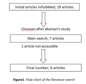
The topic of our project focuses on the anatomical variations of the dorsal motor nucleus of the vagus (DMN). Data base Pub Med was the source of our search. We inserted in advanced search of Pub Med the block chain; “anatom*” AND (“variat*” OR “categor*” OR “type*” OR “difference*” OR “version*”) AND (“human*” OR “man*”) AND (“dorsal motor nucleus” OR “DMN” OR “DMNX”[MeSH]) AND (“vagus” OR “X”). Initially, 19 articles arose. From 19, 7 of them were related to the topic of our paper and 1 article was not accessible. Consequently, for the composition of our paper 6 articles were utilized.
Discussion
The comparison of the DMN in adults and infants reveals interesting things. Although the number of neurons in the DMN remains consistent between adults and infants, adults have a higher nuclear volume and lower neuronal density in this medullary nucleus compared to infants.(3,4) Additionally, apoptosis levels in DMN neurons are higher in adults than in infants, with no significant statistical difference in glial cells. The differences in neuronal volume and density between adults and infants may be due to variations in microvascularization and distribution of neuropil. Microvascularization also plays a role in the progression of apoptosis.(5)
The DMN seems to possess and important role in Sudden Infant Death Syndrome (SIDS). Sudden Infant Death Syndrome (SIDS) is the leading cause of death in infants between the ages of one month and one year.(6) It is believed that abnormal or delayed neurogenesis in the DMN may be linked to SIDS.
Figure1. Flow chart of the literature search |
In control infants, neurogenesis in the DMN nucleus is complete at birth, with exponential postnatal growth as infants develop. However, in SIDS victims, there is a lack of development and dendritic arborization, leading to a deficiency in neuron size in the DMN. Differences have been observed in the growth of DMN neurons in SIDS infants compared to age-matched infants, with a delay in the expected loss of dendritic spines.6 Reduced neuronal density has also been observed in SIDS infants compared to normal infants.4
Immunoreactivity of the DMN. Some neurons in the DMN contain special substances such as substance-P, an undecapeptide, and tyrosine hydroxylase, a cell enzyme. Substance-P is found in both somata and fibers of approximately 16% of the total number of neurons in the DMN, totaling around 2040 neurons. The distribution of substance P positive neurons is higher in the intermediate division compared to the caudal division, with a lower presence in the rostral division. These Substance-P neurons in the DMN can appear either round or fusiform.(2) Regarding tyrosine hydroxylase; neurons that are positive for this enzyme can be round, oval, or fusiform in shape. The distribution of tyrosine hydroxylase is higher in the intermediate subdivision, while in the caudal subdivision, these neurons are only located ventrally and in the rostral subdivision, they are found medially. Notably, substance P positive neurons are primarily located in the center of the DMN, whereas tyrosine hydroxylase positive neurons are more commonly found in the periphery. Additionally, there is a difference observed in Parkinson’s disease, where substance P positive neurons are decreased in the DMN compared to non-Parkinson individuals.(2)
Conclusion
The DMN, the brainstem’s largest nucleus, is divided into sub nuclei based on functional and anatomical specialization. Despite its significance, there have been limited studies on the anatomical variations of the DMN. However, we emphasize specific anatomical differences between adults and infants, the growth pattern of neurons in SIDS infants, and the distribution of specific neuron types in the DMN sub regions. Therefore, understanding the cyto-architecture of the DMN could be essential for explaining clinical conditions and guiding future research.
References
- Huang XF, Törk I, Paxinos G. Dorsal motor nucleus of the vagus nerve: a cyto- and chemoarchitectonic study in the human. J Comp Neurol. 1993 Apr 8;330(2):158-82. doi: 10.1002/cne.903300203
- Huang XF, Paxinos G, Halasz P, McRitchie D, Törk I. Substance P- and tyrosine hydroxylase-containing neurons in the human dorsal motor nucleus of the vagus nerve. J Comp Neurol. 1993 Sep 1;335(1):109-22. doi: 10.1002/cne.903350108
- Perelló M, Cornejo MP, De Francesco PN, Fernandez G, Gautron L, Valdivia LS. The controversial role of the vagus nerve in mediating ghrelin’s actions: gut feelings and beyond. IBRO Neurosci Rep. 2022 Mar 12;12:228-239. doi: 10.1016/j.ibneur.2022.03.003
- Porzionato A, Macchi V, Parenti A, De Caro R. Morphometric analysis of infant and adult medullary nuclei through optical disector method. Anat Rec (Hoboken). 2009 Oct;292(10):1619-29. doi: 10.1002/ar.20957
- Porzionato A, Macchi V, Guidolin D, Sarasin G, Parenti A, De Caro R. Anatomic distribution of apoptosis in medulla oblongata of infants and adults. J Anat. 2008 Feb;212(2):106-13. doi: 10.1111/j.1469-7580.2007.00842.x. Epub 2007 Dec 10.
- Konrat G, Halliday G, Sullivan C, Harper C. Preliminary evidence suggesting delayed development in the hypoglossal and vagal nuclei of SIDS infants: a necropsy study. J Child Neurol. 1992 Jan;7(1):44-9. doi: 10.1177/08830738 9200700108.