Abstract
Charcot arthropathy is one of the most severe complications of diabetes and affects the quality of life of diabetic patients. Despite being the gold standard of Charcot’s arthropathy treatment, contact casting therapy requires strong adherence to the clinical pathway from both the patient and the medical personnel. We report the case of a 70-year-old male patient with a past medical history of uncontrolled Diabetes Mellitus who initially presented to the ER department of our hospital with a unilateral, swollen, warm left foot with erythema and moderate X-ray abnormalities of his left foot and ankle. He denied any previous traumatic incident. He was placed initially in a non-weight bearing contact cast. However, the patient was not compliant to his treatment and returned to the Emergency Department 4 months later with severe left foot deformity, a disarticulation of the tibiotalar and subtalar joints, and a large open ulcer of the foot. A below-knee amputation was performed. This report will therefore serve as a reminder for clinicians to keep in mind that Charcot arthropathy is a progressive condition that should be treated without delay.
Introduction
Charcot arthropathy is a major delayed complication of diabetes that affects bones, joints and the surrounding soft tissues. In the absence of normal sensation due to diabetic neuropathy, repetitive microtrauma and autonomic vascular dysfunction lead to local inflammation. Consequently, bone resorption and joint dislocation may occur.
Materials and Methods
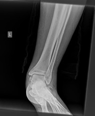
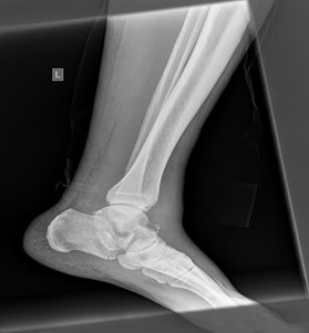
Our patient was a 70-year-old male who was admitted to our hospital with a painless, swollen, warm and erythematous left foot for 2 months. He denied any previous traumatic accident. His medical history revealed a preexisting uncontrolled Diabetes for the last 4 years. His Glycosylated Haemoglobin (Hb1Ac) level was 10,6%. Anteroposterior and lateral radiographs were obtained that confirmed the articular degenerative changes of his left ankle joint. At that moment no subluxation or any other structural deformation existed. (Figure 1 and 2). Clinical examination revealed a palpable dorsalis pedis pulse and loss of the protective sensation.
A total contact cast was chosen as a first line of treatment. The patient was discouraged from weight bearing of his left foot. Casts changes every 2 weeks for the next 4 months were recommended. However, the patient was not compliant to the therapy, and he removed the cast after three weeks. He also discarded his crutches and started to fully weight bear his limb.
Figure 1: Anteroposterior Radiograph of the ankle joint when the patient was first referred to our department |
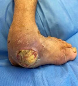
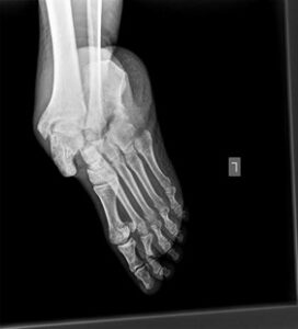
The patient presented to the Emergency Department of our hospital 4 months later for the first time since his dismissal. He had a sublaxed, swollen, erythematous foot with the presence of an open ulcer sour at the medial side of his left foot. (Figure 3). Radiographs were obtained- Anteroposterior and lateral views and a complete disarticulation of the tibiotalar and subtalar joints was confirmed. (Figures 4,5) Treatment options were discussed with the patient, and a below knee amputation was chosen as the best treatment option.
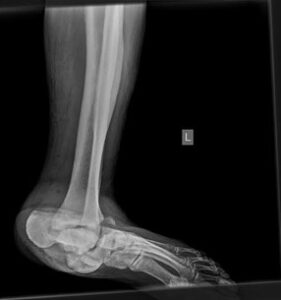
Figure 2: Lateral Radiograph of the ankle joint when the patient was first referred to our department |
Discussion
Neuropathic osteoarthropathy of the foot and ankle (Charcot foot) is a disease involving bones, joints and soft tissue of the foot that can lead to a progressive malpositioning and deformation up to complete collapse of the foot [1]. Every part of the skeleton could be affected though foot and ankle Charcot arthropathy remains the most frequent anatomic location. Most commonly, a so-called rocker-bottom deformity – a collapse of the arch in the metatarsus occurs [2]. This malalignment of the foot can cause pressure damage to the skin, open wounds, and secondary bone infection. Similarly, in our case study the patient developed very rapidly this rocket –bottom deformity which led to skin damage and open wound. (Image No 3). The presence of a rocker-bottom foot can increase the risk of a major lower extremity amputation by 15–40 times 2
Figure 3: Lateral Radiograph of the foot and ankle joint revealing dislocation and disorientation of the tibiocalcaneal, talonavicular and subtalar joints and an ulcer on the medial side |
Hastings et al have made a study looking on radiological progression of foot deformity in charcot patients by monitoring Charcot patients regularly by taking weight bearing x-rays of the foot. Their six-month data suggested worsening of medial column alignment prior to lateral column worsening [3]. This radiographic evidence of worsening foot alignment over time supports the need for aggressive intervention (conservative bracing or surgical fixation) to attempt to prevent limb-threatening complications. Salvage of Charcot neuroarthropathy complicated by a hindfoot ulcer and osteomyelitis is a complex situation. The aim of surgical intervention in an infected Charcot foot with ulceration is to eradicate the infection and obtain a stable, plantigrade foot that will allow the patient to ambulate with or without orthoses without causing any future ulcerations. Surgery in charcot foot deformities is usually recommended when infection, unstable joint and recurrent ulceration occurs. However, there is no existing protocol of what type of surgical treatment is required. Various surgical interventions have been described. A combination of talectomy and tibio-calcaneal arthrodesis was described for a Charcot foot deformity, but internal fixation was reserved for cases without foot ulcers and osteomyelitis [4,5,6]. External fixation of the midfoot prior to intramedullary fusion has also been described [7].
Figure 4: Anteroposterior Radiograph of the foot and ankle joint revealing dislocation and disorientation of the tibiotalar, talonavicular and subtalar joints |
Sohn MW et al looked at the lower extremity risk of amputation after charcot arthropathy [8]. Their results were consistent with the current practice guideline suggesting that prevention of ulceration is critical for Charcot limb salvage [9]. Their study also suggested that feet affected by Charcot arthropathy are unlikely to ulcerate when they remain clinically plantigrade and the radiographic weight-bearing relationship between the hind foot and forefoot is collinear [10,11]. These results suggest that amputation risk for Charcot arthropathy may be reduced by reserving corrective surgeries for patients with a high risk of Charcot-related ulceration.
Kucera et al, have analyzed their midterm outcomes of reconstruction of Charcot foot neuropathy in diabetic patients. A candidate for a reconstruction surgery should be a cooperating, compensated, informed diabetic patient with Charcot foot neuroarthropathy, either instable or stable, but non-plantigrade [12]. Our patient was a non-compliant non cooperative patient with a plantigrated talus, therefore a below knee amputation was thought to be the treatment of choise. The patient had a good outcome, and no complications from the wound side occurred.
Figure 5: Lateral Radiograph of the foot and ankle joint revealing dislocation and disorientation of the tibiocalcaneal, talonavicular and subtalar joints |
Conclusion
Early diagnosis and proper management, although challenging remain the most accurate prognostic factors. Treatment is based on a trial of total contact casting for early Charcot arthropathy stages with excellent results. In the presence of ulcers or skin breakdown and failure of conservative treatment operative management is indicated. Surgical intervention methods include osteotomies, internal or external fixation and amputations. His figure still awaits proper recognition.
References
- Berger-Groch J, Falkenberg CS, Schöntag S, Reize P. Case Report of an Unusual Differential Diagnosis of a Red and Atraumatic Swollen Foot Charcot Arthropathy in Rheumatoid Arthritis. J Orthop Case Rep. 2022 Nov;12(11):100-104. doi: 10.13107/jocr.2022.v12.i11.2432. PMID: 37013223; PMCID: PMC10066683.
- Marmolejo VS, Arnold JF, Ponticello M, Anderson CA. Charcot foot:Clinical clues, diagnostic strategies, and treatment principles. Am Fam Physician. 2018;97:594–9.
- Hastings MK, Johnson JE, Strube MJ, Hildebolt CF, Bohnert KL, Prior FW, Sinacore DR. Progression of foot deformity in Charcot neuropathic osteoarthropathy. J Bone Joint Surg Am. 2013 Jul 3;95(13):1206-13. doi: 10.2106/JBJS.L.00250. PMID: 23824389; PMCID: PMC3689259
- Ettinger S., Plaass C., Claassen L., Stukenborg-Colsman C., Yao D., Daniilidis K. Surgical Management of Charcot Deformity for the Foot and Ankle-Radiologic Outcome After Inter-nal/External Fixation. J. Foot Ankle Surg. 2016;55:522–528. doi: 10.1053/j.jfas.2015.12.008.
- Bajuri M.Y., Ong S.L., Das S., Mohamed I.N. Charcot Neuroarthropathy: Current Surgical Management and Update. A Systematic Review. Front. Surg. 2022;9:820826. doi: 10.3389/fsurg.2022.820826.
- Hasan K, Metikala S, Vallem MMR. Salvage of Hindfoot Charcot with Osteomyelitis and Ulceration: A Case Report. Medicines (Basel). 2022 Dec 2;9(12):61. doi: 10.3390/medicines9120061. PMID: 36547994; PMCID: PMC9781353.
- Ong SL, Bajuri MY, Mazli N. Outcome of Surgical Fixation for Midfoot Charcot Neuroarthropathy – A Systematic Review. Malays Orthop J. 2023 Mar;17(1):27-33. doi: 10.5704/MOJ.2303.004. PMID: 37064618; PMCID: PMC10103930
- Sohn MW, Stuck RM, Pinzur M, Lee TA, Budiman-Mak E. Lower-extremity amputation risk after charcot arthropathy and diabetic foot ulcer. Diabetes Care. 2010 Jan;33(1):98-100. doi: 10.2337/dc09-1497. Epub 2009 Oct 13. PMID: 19825822; PMCID: PMC2797995.
- Frykberg RG, Zgonis T, Armstrong DG, Driver VR, Giurini JM, Kravitz SR, Landsman AS, Lavery LA, Moore JC, Schuberth JM, Wukich DK, Andersen C, Vanore JV: American College of Foot and Ankle Surgeons. Diabetic foot disorders: a clinical practice guideline (2006 revision). J Foot Ankle Surg 2006; 45( Suppl. 5): S1– S66
- Bevan WP, Tomlinson MP: Radiographic measures as a predictor of ulcer formation in diabetic charcot midfoot. Foot Ankle Int 2008; 29: 568– 573
- Pinzur MS, Sostak J: Surgical stabilization of nonplantigrade Charcot arthropathy of the midfoot. Am J Orthop 2007; 36: 361– 365
- Kučera T, Grinac M, Valtr O, Šponer P. Střednědobé výsledky rekonstrukce Charcotovy neuropatické artropatie nohy u diabetiků [Mid-Term Outcomes of Reconstruction of Charcot Foot Neuroarthropathy in Diabetic Patients]. Acta Chir Orthop Traumatol Cech. 2019;86(1):51-57. Czech. PMID: 30843514.