Short Communication Platelet count as predictor of 30-day survival after intracerebral hemorrhageDespoina Avramidou, Vasileios PapadopoulosDepartment of Internal Medicine, Xanthi General Hospital, Xanthi, Greece
Correspondence Address: Vasileios Papadopoulos, MD, PhD, 2, Staliou str – 67132 Xanthi, Greece, email: vaspapmd@gmail.com
Intracerebral hemorrhage (ICH) is a major public health concern leading to high rate of mortality as well as disability [1]. Many risk factors for ICH have been
described including old age, male sex, arterial hypertension, diabetes mellitus, and high alcohol intake [2, 3]. Moreover, anticoagulants and antiplatelets have also been
documented to increase the risk for ICH [4].
The role of platelets in the pathophysiology of (ICH) has not been clarified yet. It has been demonstrated that platelet count (PLT) was significantly lower in hemorrhagic
strokes when compared with controls [5]. Moreover, PLT has been proposed as an independent predictor of poor outcome at time of discharge in cerebellar hemorrhage [6]. Interestingly,
the presence of thrombocytopenia (PLT < 150 x 103/μL) itself did not affect the functional outcome after ICH, regardless of antiplatelets administration [7]. However, whether PLT
can be a prognosticator of survival after ICH remains obscure.
We have retrospectively enrolled 60 patients, aged 75.9 ± 12.0 years, who had been admitted to Xanthi General Hospital for ICH between January 2018 and May 2021.
Demographics, medical record, vital signs, arterial blood gas test, complete blood count, blood biochemistry, and CT scan test were collected for each patient. PLT at admission (PLT1)
and 24 hours later (PLT2) were evaluated as potential predictors of 30-day survival after ICH. Descriptive statistics are provided either as means along with their relevant standard
deviations, or percentages, for scale and nominal variables respectively. Survival data are given as medians along with their 25th and 75th percentiles (Q1 and Q3, respectively).
All patients were censored, either at the time of their death, or at the 31st day after admission. Kaplan Meier (KM) curves were used to depict survival data; the log-rank test was
used to determine the univariate significance of the study variables. Cox proportional hazards regression analysis was performed to explore the potential correlations between
independent variables and survival data. All reported p values are two-sided. The level of statistical significance was set to p=0.05. All numerical values are given with at least
two significant digits. Statistical analysis was performed with the use of IBM SPSS Statistics software, version 26.0, for Windows. MedCalc software, version 20.218, was
preferred for visualization of results. The study was approved by the Scientific Board of Xanthi General Hospital (Decision No. 103/May 17, 2021).
Thirty patients (50%) succumbed within the first 30 days. The median survival time during the first month after admission was 25 days (Q1: 11 days; Q3: 30 days).
PLT1 values were 232,000 ± 86,000/μL and 211,000 ± 60,000/μL (P=0.251), while PLT2 values were 240,000 ± 63,000/μL and 203,000 ± 60,000/μL (P=0.012), for survivors
and non-survivors, respectively; these results are schematically presented as boxplots. (Figure 1)
Figure 1. Boxplots presenting PLT1 and PLT2 values in survivors and non-survivors; PLT values (y-axis) are given in 103/μL.
Using Cox proportional hazards regression univariate analysis, it has been shown that 30-day survival after ICH was positively correlated with PLT2 (P=0.012),
hemoglobin levels at admission (P=0.020), and oxygen saturation at admission (P=0.015). Moreover, 30-day survival was negatively correlated with age (P=0.001), blood glucose
levels at admission (P=0.043), medical history of diabetes mellitus (P=0.014), and medical history of arterial hypertension (P=0.036). Of note, PLT1 was comparable between
survivors and non-survivors (P=0.251).
The use of a Cox-regression proportional hazards multivariate analysis model demonstrated that increased PLT2 was independently correlated with 30-day survival after ICH,
considering all other parameters as potential confounders (HR: 0.986 per unit 103/μL; 95% CI: 0.978-0.994, P<0.001). All necessary details are provided in Table 1.
Table 1. Patients’ characteristics as well as Cox regression univariate and multivariate analysis based on 30-day survival status.
SD: Standard Deviation, † For scale and nominal variables, respectively,
‡ P-value based on univariate Cox regression analysis,
HR along with ±95% CI and P-value based on multivariate Cox regression analysis
To further assess an easy and clinically useful tool for using PLT2 as a predictor of 30-day survival, we used 225,000/μL, being the mean of the PLT2 covariate, as
derived from the Cox proportional hazards regression univariate analysis. Patients with PLT2 ≥225,000/μL had a hazard ratio (HR) of 0.991 (95% CI: 0.984-0.998) per unit (103/μL)
to succumb within the first 30 days after admission for ICH (HR<1 favors 30-day survival). The result was statistically significant (P=0.048) using the Log-Rank test; the relevant
Kaplan-Meier curve is provided as Figure 2.
Figure 2. Kaplan-Meier curve depicting survival function according to selected PLT2 cutoff; ICH patients with PLT2 ≥225,000/μL presented favorable 30-day survival
when compared to patients with PLT2 <225,000/μL (Log-Rank P=0.048).
Based on our results, we propose that PLT2 might be further investigated as an early predictor of 30-day survival after ICH. Moreover, in the light of absence of independent
correlation between PLT1 and 30-day survival, it is reasonable to hypothesize that the crucial parameter of platelet involvement in ICH might not be their initial
number per se, but rather their alterations due to vascular damage and/or activation.
References
1. Qureshi AI, Mendelow AD, Hanley DF. Intracerebral haemorrhage. Lancet. 2009 May 9;373(9675):1632-44. doi: 10.1016/S0140-6736(09)60371-8. PMID: 19427958; PMCID: PMC3138486.
2. Ariesen MJ, Claus SP, Rinkel GJ, Algra A. Risk factors for intracerebral hemorrhage in the general population: a systematic review.
Stroke. 2003 Aug;34(8):2060-5. doi: 10.1161/01.STR.0000080678.09344.8D. Epub 2003 Jul 3. PMID: 12843354.
3. Emerging Risk Factors Collaboration; Sarwar N, Gao P, Seshasai SR, Gobin R, Kaptoge S, Di Angelantonio E, Ingelsson E, Lawlor DA, Selvin E, Stampfer M, Stehouwer CD,
Lewington S, Pennells L, Thompson A, Sattar N, White IR, Ray KK, Danesh J. Diabetes mellitus, fasting blood glucose concentration, and risk of vascular disease: a collaborative
meta-analysis of 102 prospective studies. Lancet. 2010 Jun 26;375(9733):2215-22. doi: 10.1016/S0140-6736(10)60484-9. Erratum in: Lancet. 2010 Sep 18;376(9745):958. Hillage,
H L [corrected to Hillege, H L]. PMID: 20609967; PMCID: PMC2904878.
4. Zeng Z, Chen J, Qian J, Ma F, Lv M, Zhang J. Risk Factors for Anticoagulant-Associated Intracranial Hemorrhage: A Systematic Review and Meta-analysis.
Neurocrit Care. 2023 Jan 20. doi: 10.1007/s12028-022-01671-4. Epub ahead of print. PMID: 36670269.
5. Sadeghi F, Kovács S, Zsóri KS, Csiki Z, Bereczky Z, Shemirani AH. Platelet count and mean volume in acute stroke: a systematic review and meta-analysis.
Platelets. 2020 Aug 17;31(6):731-739. doi: 10.1080/09537104.2019.1680826. Epub 2019 Oct 26. PMID: 31657263.
6. Lin CY, Chang CY, Sun CH, Li TY, Chen LC, Chang ST, Wu YT. Platelet count and early outcome in patients with spontaneous cerebellar hemorrhage:
a retrospective study. PLoS One. 2015 Mar 17;10(3):e0119109. doi: 10.1371/journal.pone.0119109. PMID: 25781880; PMCID: PMC4364557.
7. Mrochen A, Sprügel MI, Gerner ST, Sembill JA, Lang S, Lücking H, Kuramatsu JB, Huttner HB. Thrombocytopenia and Clinical Outcomes in Intracerebral Hemorrhage:
A Retrospective Multicenter Cohort Study. Stroke. 2021 Jan;52(2):611-619. doi: 10.1161/STROKEAHA.120.031478. Epub 2021 Jan 12. PMID: 33430632.
Accessibility Bar
visibility_offDisable flashes
titleMark headings
settingsBackground Color
zoom_outZoom out
zoom_inZoom in
remove_circle_outlineDecrease font
add_circle_outlineIncrease font
spellcheckReadable font
brightness_highBright contrast
brightness_lowDark contrast
format_underlinedUnderline links
font_downloadMark links
Reset all optionscached
Χρησιμοποιούμε cookies για να σας προσφέρουμε την καλύτερη δυνατή εμπειρία στη σελίδα μας. Εάν συνεχίσετε να χρησιμοποιείτε τη σελίδα, θα υποθέσουμε πως είστε ικανοποιημένοι με αυτό..
This website uses cookies to improve your experience while you navigate through the website. Out of these, the cookies that are categorized as necessary are stored on your browser as they are essential for the working of basic functionalities of the website. We also use third-party cookies that help us analyze and understand how you use this website. These cookies will be stored in your browser only with your consent. You also have the option to opt-out of these cookies. But opting out of some of these cookies may affect your browsing experience.
Necessary cookies are absolutely essential for the website to function properly. These cookies ensure basic functionalities and security features of the website, anonymously.
Cookie
Duration
Description
cookielawinfo-checkbox-analytics
11 months
This cookie is set by GDPR Cookie Consent plugin. The cookie is used to store the user consent for the cookies in the category "Analytics".
cookielawinfo-checkbox-functional
11 months
The cookie is set by GDPR cookie consent to record the user consent for the cookies in the category "Functional".
cookielawinfo-checkbox-necessary
11 months
This cookie is set by GDPR Cookie Consent plugin. The cookies is used to store the user consent for the cookies in the category "Necessary".
cookielawinfo-checkbox-others
11 months
This cookie is set by GDPR Cookie Consent plugin. The cookie is used to store the user consent for the cookies in the category "Other.
cookielawinfo-checkbox-performance
11 months
This cookie is set by GDPR Cookie Consent plugin. The cookie is used to store the user consent for the cookies in the category "Performance".
viewed_cookie_policy
11 months
The cookie is set by the GDPR Cookie Consent plugin and is used to store whether or not user has consented to the use of cookies. It does not store any personal data.
Functional cookies help to perform certain functionalities like sharing the content of the website on social media platforms, collect feedbacks, and other third-party features.
Performance cookies are used to understand and analyze the key performance indexes of the website which helps in delivering a better user experience for the visitors.
Analytical cookies are used to understand how visitors interact with the website. These cookies help provide information on metrics the number of visitors, bounce rate, traffic source, etc.
Advertisement cookies are used to provide visitors with relevant ads and marketing campaigns. These cookies track visitors across websites and collect information to provide customized ads.
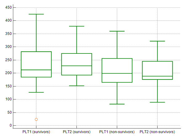 Figure 1. Boxplots presenting PLT1 and PLT2 values in survivors and non-survivors; PLT values (y-axis) are given in 103/μL.
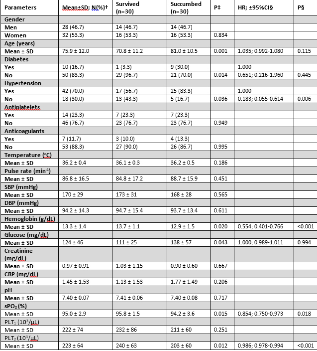
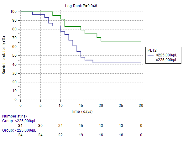 Figure 2. Kaplan-Meier curve depicting survival function according to selected PLT2 cutoff; ICH patients with PLT2 ≥225,000/μL presented favorable 30-day survival
when compared to patients with PLT2 <225,000/μL (Log-Rank P=0.048).
Based on our results, we propose that PLT2 might be further investigated as an early predictor of 30-day survival after ICH. Moreover, in the light of absence of independent
correlation between PLT1 and 30-day survival, it is reasonable to hypothesize that the crucial parameter of platelet involvement in ICH might not be their initial
number per se, but rather their alterations due to vascular damage and/or activation.
Figure 2. Kaplan-Meier curve depicting survival function according to selected PLT2 cutoff; ICH patients with PLT2 ≥225,000/μL presented favorable 30-day survival
when compared to patients with PLT2 <225,000/μL (Log-Rank P=0.048).
Based on our results, we propose that PLT2 might be further investigated as an early predictor of 30-day survival after ICH. Moreover, in the light of absence of independent
correlation between PLT1 and 30-day survival, it is reasonable to hypothesize that the crucial parameter of platelet involvement in ICH might not be their initial
number per se, but rather their alterations due to vascular damage and/or activation.